by Whitecoat
The Journal of the American College of Cardiology presented the ROSE study for triaging patients with syncope in the emergency department. No, ROSE isn’t some LOL that the study was named after. ROSE is an acronym standing for “Risk Stratification of Syncope in the Emergency Department.” They just left out a few letters because an acronym of “RSOSITED” just isn’t quite as catchy. Maybe SOS-ED would have been cooler, but ROSE it is.
Anyway, the study looked at what factors were likely to be present in patients who passed out and who had a “serious outcome” or death in the following month. Serious outcomes or death occurred in 7% of all patients who passed out in this study. They found that positive fecal occult blood, low hemoglobin levels, low oxygen saturation, and Q waves on the EKG were all predictive of worse prognosis for patients with syncope.
In addition, a BNP (brain natriuretic peptide) level greater than 300 was present in 36% of syncopal patients who later suffered serious cardiovascular events and in 89% of syncopal patients who later died.
More than 98% of patients who had none of these risk factors also had no serious outcome or death in the subsequent month after their syncopal event.
So check the BNP on syncope patients and get out those rubber gloves, ladies and gents. Add syncope to the list of patient complaints for which rectal exams may be indicated.
After all this, if you’re still wondering what “LOL who DFO” means, then you have to read this religious post.
Thursday, July 29, 2010
Do Doctors Rat Out Their Own?
Do doctors rat out their own? That's an interesting question. A reader sent me this picture of newspaper text with a similar title. I did a little search and found the source study for this story was a JAMA article that studied the ratting out habits of almost 2000 physicians. What did they find? Almost 2/3 agreed they had a professional obligation to report incompetent physicians or physicians under the influence of drugs or alcohol.
Only 17% (about 400) physicians reported having direct knowledge of an incompetent or intoxicated physician and of those 17%, 2/3 (or about 250) of these physicians reported them to appropriate authorities. That leaves only about 150 out of 5000 physicians, or 6% of physicians who have not reported their colleagues when they should have.
Why did this 6% of physicians not report incompetent or intoxicated physicians? The most common reasons reported were

- Fear of retribution
- It was being handled by someone else
- Nothing would happen anyway
- The physician would be excessively punished
- It wasn't their responsibility
All the Google headlines seem to say no, physicians would not rat out their own. The power is in perspective. What if the headline said "94% of physicians acted appropriately" instead of "Physicians don't rat out their own"
I look at the data and find physicians as a whole carry the highest degree professionalism, compassion and competency. How many Wall street executives were reporting their colleagues while the country was looted? How many friends and colleagues turn in their mother, son or neighbor falsifying their disability claims or their mortgage applications? How many Obama officials are failing to tell the public the truth as they recapitalize banks with record profits at the expense of fixed income elderly. How many SEC executives were looking the other way when their colleagues were surfing porn eight hours a day. How many Congressmen and women ignore their colleagues' flagrant dishonesty by buying votes from special interests. How many cops let one slide for their brother in the badge.
Every where you look, you are going to have people who lack the courage to do the right thing. If you believe that physicians are somehow super human and will always turn their colleagues in you might as well write a letter to the tooth fairy.
What you have here is a tiny part of a tiny problem. Physicians as a whole are highly competent. They have passed the test. They have paid thousands of dollars to certify with their governing boards to declare their competency with certification. If we are now to believe that physicians are incompetent despite their certification by their specialty board, we then must abandon the model of board certification and find an alternative means to measure the competency of physicians.
And when I am forced to pay money and reapply for state licensure every few years, my colleagues are forced to attest (in uncompensated time) as to the competency of my skills and the lack of my addiction to drugs and alcohol. If physicians are practicing drunk, and the state licensure process is meant to prevent it, then the government process is flawed, not the physicians. The process relies on physicians providing truthful answers and physicians as a whole, 94% of them, are truthful. If the concern is missing 6% of the problem then the government process should be changed. Just like you can't change your spouse, you're not going to change the moral fabric of the physician.
I'll take a 94% success rate any day of the week. The other 6% will eventually be found, one way or another. Besides, there's always a good med mal lawyer or two or three waiting for an opportunity to protect the patient.
Wednesday, July 28, 2010
Video: Awake Endotracheal Intubation
Video: Awake Endotracheal Intubation for "Fun and Knowledge" by an anesthesiologist from the Massachusetts General Hospital (MGH). Not sure about the "fun" part...
References:
Awake Endotracheal Intubation for Fun and Knowledge. Medgadget, 2010.
Posted at Clinical Cases and Images. Stay updated and subscribe, follow us on Twitter and connect on Facebook.
Tuesday, July 27, 2010
The art of medicine and whether computers can replace doctors in Diagnosis and treatment
by Eric Van De Graaff, MD
My medical school at the University of Utah developed a clever computer program in the late 80s that was meant to both educate medical students and assist in the treatment of patients.
For several years the tech geeks at the school had collected an immense database of information from all the patient admissions at the hospital—presenting symptoms, exam findings, tests, and final diagnoses. They took all this data and crunched it into a program that provided a statistical snapshot of all the clinical syndromes seen by our university hospital over several years and then created a user-friendly interface that allowed us new trainees to learn from all this experience.
With this program I was able to enter the term “chest pain” and quickly learn what the ultimate diagnosis was for all the thousands of patients who had reported chest pain as part of their initial symptom constellation (e.g. 12% heart attack, 6% pulmonary embolus, 46% esophageal reflux, 18% chest wall pain, etc.).
Even cooler was the function that allowed us to enter several symptoms and findings and the computer would spit out the most likely diagnosis. Mind you, this was not based on some sort of theoretical textbook list of syndromes, but rather on the real-world experience of our patients and our doctors at our hospital. I’d type in “abdominal pain, nausea, and elevated blood lipase” and the computer would tell me that this patient was 91% likely to have acute pancreatitis.
As far as a computerized training tool this program was unsurpassed. It could tap its database to create a fictionalized patient and problem list, then quiz us on what testing and therapy we’d recommend. Patient X with foot pain and history of diabetes, heart disease, and schizophrenia shows up with a fever. What’s your next test? What’s the most likely diagnosis?
As medical students, we couldn’t help but be a little intimidated by a computer program that seemed a million times smarter than we were. It seemed to us that the rapidly advancing technology, if left to evolve down this Orwellian path, would soon be able to diagnose and treat patients with precision and reliability that we humans could only dream of. It was a little deflating to think that we would suffer through a decade of schooling and countless thousands in loans only to find that we are all being replaced by computers.
Well, that was 1989 and in the 21 intervening years I’ve managed to find employment as a doctor and have not yet been replaced by a robot. Despite the fact that we have more evidence-based models of care (algorithms based on large population studies that help take the variability out of medical care) than we’ve ever had, human doctors—despite all their flaws and imperfections—are still an integral part of modern healthcare.
You wouldn’t think this should be the case. In a world where complex software at sites like Amazon and Pandora can predict your tastes better than you can, where Google can access trillion pieces of information in fractions of a second, where all details of everyone’s medical history will potentially be available for data mining, and where computers can trounce the grandmasters in chess, someone could come up with a computer program that would out-doctor even the best of doctors (is there an app for that?). How is it that our system continues to rely on the imperfect judgment of doctors to treat us?
The answer, I would suggest, is what we commonly know as the “art of medicine.” Most of you have heard this phrase before, but do you know what it actually means?
I don’t either. Look it up in a dictionary and you’ll come up empty. Wikipedia, one of my favorite resources, has no separate entry for the art of medicine. One of the few on-line essays I found dealing with the subject employed vague platitudes to define this concept: “Mastery. Individuality. Humanity. Morality.”
I would attempt to define “the art of medicine” a little more specifically.
In much of modern medicine there are pretty clearly established practice patterns that lead to reliable results. A good example of this is our treatment of acute appendicitis. Ever wonder why there aren’t a half dozen competing treatment strategies for appendicitis? Come down with leukemia and you’ll be presented with at least a couple of options for therapy (different types of chemotherapy, bone marrow transplant, alternative medicines, etc.), but get an infected appendix and you pretty much get an immediate trip to the operating room. The simple reason you don’t get many options with appendicitis is that the established therapy works so well—it’s curative in all but rare cases. The rate of cure for most leukemias is low, thus there are many different ways to produce a relatively poor outcome. If we had a pill that cured leukemia in nearly all cases, all other alternatives would quickly vanish.
The treatment of appendicitis is so straightforward (the diagnosis, by the way, is murkier) that even the simplest computer program would churn out the correct recommendation every time. One plus one equals two. At that point all you need is a pair of hands to dig the offending tissue out of the abdomen.
But what happens when one plus one doesn’t equal two? What happens when there is no cure, or when the intended cure doesn’t produce the results you hope for, or when a cure is available but the patient’s condition precludes its application? I maintain that the art of medicine is very simply defined as what you do when things don’t go how they’re supposed to—when one plus one adds up to something else.
The art of medicine is what you lean on when you have to explain to family members why the surgery you just performed on their loved one didn’t produce the beneficial results you had hoped for, or, even worse, resulted in an adverse outcome or complication.
The art of medicine guides you in dealing with an emotionally fragile patient who needs a procedure or treatment that you know will be beyond their ability to cope.
The art of medicine dictates how you break the news to a worried waiting room that their ill family member has just passed away; or how to cautiously guide families to the acceptance of impending demise in a patient about whom they clearly have denial; or how to comfort a surviving spouse when he or she asks you if they should have done more to save the person they’d been married to for 50 years.
The art of medicine is knowing when to stop asking questions during an office visit and just let the patient speak what’s on his or her mind. It’s knowing when the agenda you have for your interaction has to take a back seat to something that may be very unimportant to you but critical in the mind of your patient.
The art of medicine is how you handle the patient who returns faithfully to your office seeking relief of her symptoms but who just can’t bring herself to remain compliant with the medications you prescribe to treat her condition. Or how you handle the patient with an easily treatable illness who simply refuses therapy.
The art of medicine is choosing a course of therapy based as much on an understanding of the character and personality of the patient as on knowledge the disease process itself.
I could go on for page after page but I think you get the picture. To a computer program—even the most clever ones—one plus one will always be two and the treatment of medical problems will remain nothing more than a function of odds, statistics, and search engines. This may be satisfactory for appendicitis and a handful of other problems, but for most of what we do as doctors the math is never quite so simple.
The art of medicine—the art of dealing with the unanticipated, unwanted, and less-than-optimal—can’t be programmed into a computer and, for that matter, can’t be taught in medical school. It’s what clinicians develop after years of experience, application and sometimes failure of science, mistakes, introspection, and learned humility.
Those sorts of skills will be in high demand forever. And until they can program robots to do all that I’ll probably be able to keep my job.
Eric Van De Graaff is a cardiologist at Alegent Health who blogs at the Alegent Health Cardiology Blog.
Lost in Translation
July 28, 2010 By Mike Cadogan (life in fast lane)
Whether it is interpreting a physicians handwriting, transcribing the muffled rants of the dictating clinician or battling the auto-correct function of microsoft word…meaning can sometimes be lost in translation…
The patient has no previous history of suicides
Patient has left her white blood cells at another hospital.
Patient’s medical history has been remarkably insignificant with only a 40 pound weight gain in the past three days.
She has no rigors or shaking chills, but her husband states she was very hot in bed last night.
Patient has chest pain if she lies on her left side for over a year.
On the second day the knee was better and on the third day it disappeared.
The patient is tearful and crying constantly. She also appears to be depressed.
The patient has been depressed since she began seeing me in 1993.
Discharge status:- Alive, but without my permission.
Healthy appearing decrepit 69-year old male, mentally alert, but forgetful.
Patient had waffles for breakfast and anorexia for lunch.
She is numb from her toes down.
While in ER, she was examined, x-rated and sent home.
The skin was moist and dry.
Occasional, constant infrequent headaches.
Patient was alert and unresponsive.
Rectal examination revealed a normal size thyroid.
She stated that she had been constipated for most of her life until she got a divorce.
I saw your patient today, who is still under our care for physical therapy.
Both breasts are equal and reactive to light and accommodation.
Examination of genitalia reveals that he is circus sized.
The lab test indicated abnormal lover function.
Skin: somewhat pale, but present.
The pelvic exam will be done later on the floor.
Large brown stool ambulating in the hall.
Patient has two teenage children, but no other abnormalities
When she fainted, her eyes rolled around the room.
The patient was in his usual state of good health until his airplane ran out of fuel and crashed.
Between you and me, we ought to be able to get this lady pregnant.
She slipped on the ice and apparently her legs went in separate directions in early December.
Patient was seen in consultation by Dr. Smith, who felt we should sit on the abdomen and I agree.
The patient was to have a bowel resection. However, he took a job as a stock broker instead.
By the time he was admitted, his rapid heart had stopped, and he was feeling better.

Never take an X-ray request literally...
Defensive medicine forces residents to use test oriented care
by a medical resident
A recent article in US News and World Report, Most U.S. Physicians Practicing Defensive Medicine, claims that physicians are ordering more tests and escalating the work-up of sick patients, all in the name of defensive medicine.
Is it true? Absolutely.
It is especially true in the emergency department, where we have one shot to get things right, or else. Ironically, by practicing defensive medicine we are not practicing quality medicine – and isn’t that what patients really want?
Training during residency has changed over the last two decades, and the emphasis has shifted from performing a good history and physical examination to ordering and interpreting laboratory tests and imaging studies. We no longer take the time to listen to our patients. Instead, we have started clicking as many buttons on the computer order set as we possibly can in order to cover every life-threatening diagnosis. But even in the world of technology, numbers, and images, medicine remains an imperfect science.
I vividly remember a lecture during medical school on cardiology. The professor, who was in his 80s at the time, taught us how to use the stethoscope and explained the subtleties of different heart sounds. He gave us a CD, which is now sitting somewhere in my closet under a film of dust. There is an art to distinguishing a rub, ejection murmur, opening snap, split S1, gallop, click, or extra heart sound – especially when the sounds are so soft that they can barely be heard in a completely silent room.
It takes extensive practice and training to improve at this – but rather than using our stethoscope, we spend time looking at echocardiograms and troponin levels. As another example, there have been several articles written on how poorly residents and attendings perform a neurologic exam in the emergency department – a good neurologic exam takes time and requires the full, undivided attention of a perceptive clinician. Some people have a knack for this and others do not – regardless, physician training in these areas is far from what it used to be. Over the last year, I can count on one hand the number of times I have been accompanied at the bedside by an attending who lays his hands on the patient.
Our predecessors were able to gather essential pieces of clinical data from a physical exam. Today, in the world of overburdened emergency departments, full hospitals, and electronic ordering and note-writing systems, we are forced to spend less and less time with our patients. In an attempt to compensate for this problem, we make up in quantity what we cannot provide in quality – and we make up with money what we cannot provide in time. Although the perception is that patients benefit, by getting a myriad of lab tests and imaging studies, they do not. These tests mean very little unless they are correlated clinically. They only become significant in the setting of the patient.
Rather than realizing this, clinicians have begun to practice test-centered medicine rather than patient-centered medicine. This causes huge delays and expenses in patient care. It also places patient at risk for, 1) being treated unnecessarily for incidental findings; and, 2) being exposed to unnecessary radiation. Furthermore, it alienates patients even further from their physicians – and this, perhaps, is the greatest cause of increased lawsuits and patient dissatisfaction, which starts the cycle of practicing defensive medicine all over again.
This anonymous medical resident blogs at A Medical Resident’s Journey.
Takotsubo cardiomyopathy (broken-heart syndrome) in differential diagnosis of chest pain
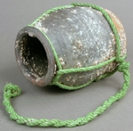
Takotsubo cardiomyopathy (also called stress induced cardiomyopathy, apical ballooning, or broken heart syndrome) was first described in Japan 20 years ago. It is typically precipitated by acute emotional stress, hence the names “stress cardiomyopathy” or “broken-heart syndrome.”
Takotsubo cardiomyopathy is characterised by acute, reversible left ventricular dysfunction in a distribution,which does not correlate with the coronary artery blood supply. The left ventricular dysfunction occurs without obstructive coronary artery disease (CAD) and usually resolves spontaneously over a period of weeks.
The characteristic appearances on contrast angiography include:
- ballooned apical segment
- hypercontractile basal portion of the left ventricle
The appearances are reminiscent of the design of the traditional fishing pot used in Japan to trap octopus, hence the descriptive term "tako-tsubo" cardiomyopathy (octopus trap, tako tsubo). Such a trap, no more than simple ceramic jar, take advantage of the octopus’ preference for small, enclosed spaces and the security they seem to promise. They are simply left on the seabed and gathered later after octopi have had time to occupy them.
Although Takotsubo cardiomyopathy was initially considered rare, it could possibly be responsible for 1-2% of admissions for acute coronary syndrome in industrialised countries.
Although Takotsubo cardiomyopathy was initially considered rare, it could possibly be responsible for 1-2% of admissions for acute coronary syndrome in industrialised countries.
Takotsubo Cardiomyopathy, or Broken-Heart Syndrome. SS Virani et al, Tex Heart Inst J. 2007; 34(1): 76–79.
Image source: Octopus trap, tako tsubo, Morikami museum.
Monday, July 26, 2010
Article Review: Premature diagnostic closure

You are taking care of a patient, who frequently presents to the ED for polysubstance use. You are pretty sure his altered mental status is from polysubstance use again. He was found in his home next to drug paraphernalia. He intermittently becomes severely agitated, and so you give him sedatives. He has a low-grade fever, but you attribute that to his psychomotor agitation and likely stimulant use. Because he remains confused and lethargic after 8 hours, you admit him to an inpatient team to await further metabolism of his recreational drugs and your sedation medications.
The next day, you learn that had meningoencephalitis.

Cognitive biases are often the root cause for medical errors.
Specifically premature closure is the #1 cause of diagnostic errors. This Academic Medicine article attempts to study this concept of physician diagnostic flexibility (changing one's mind about the patient's diagnosis during the case presentation). Is it just a matter of teaching learners to avoid premature closure?
Specifically premature closure is the #1 cause of diagnostic errors. This Academic Medicine article attempts to study this concept of physician diagnostic flexibility (changing one's mind about the patient's diagnosis during the case presentation). Is it just a matter of teaching learners to avoid premature closure?
Methodology
- 256 primary care physicians viewed a simulated patient vignette video.
- Physicians were divided into MORE experienced (attended medical school during 1960-1987) and LESS experienced physicians (attended medical school during 1996-2001).
- The video is of a patient with signs and symptoms consistent with coronary heart disease (CHD). For the sake of authenticity, these patients concurrently exhibited some GI and stress-related symptoms.
- At the mid-point of the case, the physicians were surveyed about their initial impressions of the case.
- At the end of the case, the physicians were surveyed about their final diagnosis and disposition plans.
 ResultsThe results actually were expectedly quite confusing. There are multifactorial causes for diagnostic flexibility and premature closure.
Case mid-point: More experienced physicians diagnosed CHD correctly (66%) compared to less experienced physicians (55%). This is no surprise. With more experience, you may make the correct diagnosis sooner.
Physicians who were the most likely to change their minds:
- Those with LESS experience. Specifically, if less experienced physicians selected a CHD diagnosis at the mid-point of the case, they were more likely to shift to a non-CHD diagnosis by the end of the case, compared to more experienced physicians. In this case, diagnostic flexibility was an undesired outcome. Interestingly, both less and more experienced physicians changed their diagnosis from non-CHD (mid-point) to a CHD final diagnosis with about equal frequency.
- Those who named a non-CHD diagnosis as their mid-point impression.
- Those who did not ask about patient's prior cardiac disease.
 So what's the take-home point?
- Clinical experience is invaluable.
- Experience is likely more important than merely teaching learners to reason with a more analytical approach to avoid cognitive errors.
ReferencesEva KW, Link CL, Lutfey KE, & McKinlay JB (2010). Swapping horses midstream: factors related to physicians' changing their minds about a diagnosis. Academic medicine : journal of the Association of American Medical Colleges, 85 (7), 1112-7 PMID: 20592506
Bronchospastic Blood Pressure Badness
shared from http://lifeinthefastlane.com
aka Pulmonary Puzzle 013
You arrive at work early and notice a considerable commotion in the resus area of the emergency department. A nurse spots you, and waves at you to come over. The medical team, at the end of their night shift, are stressed, sleep deprived and look worried.
A critically ill young man is now hypotensive following intubation. He was intubated for a severe asthma attack resulting in type 1 and 2 respiratory failure. The team leader asks you for help.
Questions
Q1. What are the most important things to check when there is a problem with a mechanically ventilated patient? You may remember this question from Pulmonary Puzzle 012 – Man versus Machine — it is repeated for a reason… It’s important!
Q2. What is the most important first step in managing the patient who is hypotensive soon after intubation?First determine the severity of the problem — do you need to start immediate resuscitation?Then assess MASH:
Now you can attempt to diagnose the problem.
- Movement of the chest during ventilation —
is it absent or is movement only on one side? Is the chest hyper-expanded?- Arterial saturation (SpO2) and PaO2 —
obtain an ABG sample- Skin colour of the patient (is he turning blue or pinking up?) —
the SpO2 monitor lags behind the true oxygen saturation of the patient.- Hemodynamic stability.
Disconnect the the endotracheal tube from the ventilator circuit.In asthmatics, this may be life-saving. If the cause is dynamic hyperinflation (‘gas trapping’) blood pressure will rise over 10-30 seconds as the gas is released.
In general, the causes of hypotension or shock are PRROVV:
Q4. What are the other early management priorities?- Cardiogenic
-
- Pump (e.g. imparied contractility, valve dysfunction)
- Rate (fast or slow or absent)
- Rhythm (regular or irregular)
- Obstructive (e.g. tension pneumothorax, pericardial tamponade, PE, dynamic hyperinflation)
- Vasodilation = distributive shock (e.g. sepsis, anaphlaxis, neurogenic, hepatic failure)
- Volume depletion = hypovolemia (e.g. dehydration, hemorrhage, third spacing)
- Hypovolemia exacerbated by decreased venous return due to positive intrathoracic pressure.
- Vasodilation and myocardial depression due to the induction drugs used for rapid sequence intubation (e.g. thiopentone, propofol).
- Dynamic hyperinflation (gas-trapping) due to excessive ventilation — especially in the patient with bronchospasm.
- Tension pneumothorax due to positive-pressure ventilation.
The patient has already been disconnected from the ventilator circuit.Important management priorities include:
- Administer high-flow oxygen (FiO2) via a bag-valve-mask and manually ventilate (usually <10 breaths/min) following adequate disconnection to allow the release of trapped gas.
- Consider needle thoracostomy for tension pneumothorax — carefully consider whether there is time for confirmation by bedside ultrasound or chest x-ray (there often is), so that an unnecessary invasive procedure is not performed. If the chest is needled, formal intercostal catheter insertion is mandatory.
- Administer 10-20 mL/kg IV fluid boluses to overcome the cardiovascular effects of induction drugs and/or unmasked hypovolemia. Vasopressors (e.g. metaraminol 0.5-1mg IV boluses) may also need to be administered as a temporizing measure.
Never intubate an asthmatic… unless you absolutely have to!Intubation and ventilation may be life-saving, but carries significant risks. There are the usual risks such as failed intubation, airway trauma, aspiration, and increased risk of stress ulceration and nosocomial pneumonia. But there are additional risks specific to the patient with reactive airways disease.
These include:
- inadvertent pulmonary hyperinflation.
- hypotension
- barotrauma and pneumothoraces
- PEA arrest due to dynamic hyperinflation.
- aggravation of bronchospasm.
- longer term risk of myopathy from the combination of corticosteroids and neuromuscular blockade required to facilitate mechanical ventilation.
- cardiac or respiratory arrest
- severe hypoxia (e.g. hypoxic seizure)
- rapidly deteriorating level of consciousness
These relative indications need to be balanced against the risks of intubation. Hyperacute asthma may have hypercapnea due to mechanical limitation of ventilation rather than fatigue, and this may improve with aggressive treatment.
- progressive patient fatigue
- hypercapnea
There is no clear evidence for the superiority of one ventilation mode over another (i.e. volume-controlled versus pressure-controlled).Initial ventilator settings (volume-controlled ventilation):
- Tidal volume 6-8 mL/kg
- Slow respiratory rate (e.g 8-10/min)
- High inspiratory flow rate (e.g 80-100L/min) to allow longer expiratory times
- PEEP of 0 cmH2O (some experts like a bit of PEEP — more on that another time…)
- FiO2 titrated to keep SaO2 >93%.
Expect the following with these initial settings in a patient with asthma:
- high peak inspiratory pressures (PIP) — don’t worry this does not necessarily correlate with lung barotrauma.
- respiratory acidosis due to a low target minute ventilation — sedation and neuromuscular blockade may be required to suppress spontaneous ventilation.
References
- Bersten AD, Soni N. Oh’s Intensive Care Manual (6th edition). Butterworth-Heinemann, 2008.
- Gomersall C. ICU Web — Trouble-shooting mechanical ventilation
- Life in the Fast Lane. Acute severe asthma
- Life in the Fast Lane — ICU Mind Maps: Mechanical Ventilation; Haemodynamic Effects of Ventilation
- Weingart S. EMCrit Lecture – Dominating the Vent: Part II; EMCrit Podcast 15 – the Severe Asthmatic; EMCrit Podcast 16 – Coding Asthmatic, DOPES, & Finger Thoracostomy
Blast From The Past Post From 2008.
Here's an old post of mine on a slow Sunday morning. I was reminded of this oldy but goody after a reader left a comment on this 2008 post of mine explaining why the mainstream media hates doctors so much. Go read the post on why the mainstream media hates doctors so much. I'll give you the gist of the post. Doctors are hated because they work with poor people and poor people see doctors as being rich. All other professionals with post graduate education degrees work mostly with other rich people, so they are out of site and out of mind to the poor. When was the last time you heard the media complaining about all those rich successful architects or small business men and women?
Go read the post, then read this comment left by a medical student. These are your future doctors. They are emotionally spent and they haven't even experienced real life medicine yet. Think they'll work for free after 25 years of continuous education? Think again.
Medical Student:
I am the son of a physician (family doctor) and I must say that I never really understood why people considered doctors to be wealthy. However, I did end up buying into it, yet I then came to the conclusion that maybe we were "wealthy" compared to the guy that works at McDonald's, Target, Blockbuster, etc. However, when I read your blog I realized why it is true that people, wrongfully so, consider doctors to be wealthy. I completely agree with the statement that doctors are compared to the poor and should not be. Doctors should be compared to lawyers, CEOs, stock brokers (a few of my friends went into this field and even during the "rough times" they are still making 200K!!!!) and other professionals. I just looked up to see when the last post was and realized that nobody is probably reading this blog anymore, I wonder how much hell you got for posting this from those that knew you. An MBA or CEO goes out and says we need to make more money, and those working for him cheer him on as a great leader and business man, a doctor tells the nurses and his staff that and they look at him as the greedy guy whom is already making more than they do and should be happy with it. I agree, you get what you pay for. Also, you don't have to accept Medicare and Medicaid (which I personally will not). If other physicians plan on accepting it, so be it. I'm sure it will be rough to turn down people, but that's what I'll pay my staff and office manager for, if not, I'll find another one that will do it, besides let us remember, we are trying to run a business. If you're a doctor and want to work for Medicare or Medicaid pay then go right ahead, don't give me your crap about we're doing this to help people, go and work for free if that's what gets you off. What other field can you say, hey you know what, I think its nice that you are charging 10 dollars for this, but I think I will only pay 3 dollars, thanks. By the way, if I end up using your product and hurt myself, I'm going to sue you. I wish you would keep posting although I'm sure there are a lot of problems with physicians that feel the same way you do posting a blog like this. Such is life right? Since I've posted this sort of commentary in the past and later on I'll get a reply saying something about how much pain and suffering there is in Africa, the Middle East, and other parts of the world, let us keep in mind what the author of this blog was trying to discuss, compare apples to apples. Besides if you want to look for the poor you don't have to go all the way to Africa, just downtown.
http://dragon666-mypictures.blogspot.com/
Sunday, July 25, 2010
Paucis Verbis card:Rapid sequence Intubation

The key to success in performing procedures is preparation. This is especially true for endotracheal intubations in the Emergency Department where things are chaotic. Strategic planning and anticipation of obstacles during rapid sequence intubation (RSI) are key principles to avoiding complications.

Do you have any good tips or mnemonics?
Feel free to download this card and print on a 4'' x 6'' index card.
Paucis Verbis card: Urine Toxicology Screen

In the Emergency Department, we often order urine toxicology screens for patients with altered mental status without an obvious cause. I find that patients are often rather forthcoming about their drug use, if they are alert enough to talk. In those cases, ordering a urine toxicology screen is unnecessary.

When you do order a tox screen, however, how do you interpret the information? While the results is a binary answer (positive vs negative), there are some nuances to interpretation. For instance, how long does a patient with urine toxicology remain positive for the drugs? Are there any medications that can cause false positives? See the helpful table below from a great review article in American Family Physician.
Check out what your laboratory screens for and, more importantly, what it does NOT screen for. Our lab, for example, does not screen for PCP but does screen for MDMA (ecstacy). That isn't a big deal, since patients who ingest PCP aren't too hard to detect clinically. They have crazy vertical nystagmus, and often there are at least 6 police officers trying to restrain the yelling patient.

Feel free to download this card and print on a 4'' x 6'' index card.
Standridge JB, Adams SM, & Zotos AP (2010). Urine drug screening: a valuable office procedure. American family physician, 81 (5), 635-40 PMID: 20187600
Dr Hysterical and Company

In a code/critical situation it takes all kinds. Here are some of the people you wish WEREN'T there....
1) Spaz - acts like they know what they are doing, but create chaos in a critical situation.
2) Hero (aka Cowboy) - run to be in on a critical even if it isn't their patient.
3) Scardy cat - they are assigned to the critical rooms, but plant themselves at the computer to admit the patient, do meds, while everyone else does their work for them.
4) Shaky Shirley - scared shitless, often new, deer in the headlights look.
5) Superior Stan - when someones fumbles around with a piece of equipment that is not often used, rolls their eyes and says: "let me show you..."
6) Dr hysteric - when their patient goes bad their voice goes up several octaves and is like fingernails on a chalkboard.
7) Dr Lazy - watches the nurses try to start an IV on a patient 10 times instead of putting in a FRICKIN' CENTRAL LINE! Hellllllooooooo!!! Or waits til the 02 sats are in the 50's to INTUBATE!
8) Save 'em all Susie - puts a 99 year old through an hour code and all that goes with it. Won't let people die even when they really want to.
9) Neck craners - the type of person who slows traffic to look at an accident scene. Crowds into the room with a critical to stand around and watch until someone tells them to GET THE F-- out! if you don't belong here. When a code is brought to the ER from somewhere in the hospital these people come in droves.
10) Mad max - doctor who takes on an angry demeanor in a code telling people to hurry it up and do what he/she says. Yells at people. Cusses. Generally pisses off the staff.
emergency room nurse
Saturday, July 24, 2010
Good medicine sometimes makes patients unhappy
shared from Kevinmd.com
As physicians, we all strive to practice good medicine. Good medicine means evidence based medicine in the patient’s best interests. In the ideal world this will make patients happy and satisfied. If you are getting the best treatment for your condition you should be happy, right?
In the real world, though, keeping patients or their families’ happy and practicing good medicine might not be possible at the same time. This is true for both inpatient and outpatient physicians.
A recent experience that one of my partners had to go through demonstrates the point. The patient in his mid 80’s came with a massive heart attack. He had a heart attack at home and, unfortunately, wasn’t found until later. He developed muscle breakdown that affected his kidneys. He had to be started on continuous dialysis.
Despite aggressive medical management, his condition had progressively deteriorated. The blood pressure remained low despite the high doses of medications. All major organs started to shut down. The patient was dying.
When his condition suddenly deteriorated and he developed a fatal arrhythmia, the responding physician refused to escalate care and suggested to the family that comfort care was more appropriate in his case.
The family was unable to make a decision, insisting on providing futile care. Subsequently, they became angry with the physician and complained to the hospital administration. This caused the physician emotional distress and an unnecessary headache. The refusal to provide futile care lead to a very unhappy family yet it was the right thing to do. It was the right thing for the patient.
Things might not be as dramatic in the outpatient world, yet the problem, probably, exists on an even bigger scale. Studies have shown that physicians are more likely to prescribe medications and order tests when confronted with a specific request from the patient. Often the request is granted even though it might not be the best treatment for the patient. Some studies have shown that the perception of the quality of care improves once the request is granted.
Some hospitals and clinics are even trying to improve patient satisfaction scores by adjusting the physician’s compensation and bonuses based on the patient satisfaction. Does that encourage physicians to do what the patient wants and not what the patient really needs?
A study published in the Archives of Internal Medicine demonstrated that the request for antidepressant prescription is much more likely to be granted if the patient asks for the medication directly or indirectly. In many cases these prescriptions would be considered unnecessary or even inappropriate by the current practice guidelines.
Any physician ever practicing outpatient primary care knows that patients often expect to be given antibiotics for upper respiratory symptoms, even though, viral infection is the culprit in more than 90% of cases. You might say: “What’s a big deal if the patient takes antibiotics for a few days? Even if unnecessary, it might make the patient feel like he is actually being treated.”
Now, imagine on the national level how much wasteful cost it adds to medical care. The patients are being exposed to unnecessary risks of antibiotics. Antimicrobial sensitivity will be altered in the community with emergence of drug resistant pathogens.
The bottom line is – practicing good medicine and having satisfied patients often means performing a balancing act on the part of inpatient and outpatient physicians. The silver lining, according to the study mentioned earlier, is that effective communication with the patient is shown to improve satisfaction even when the specific requests are not being granted.
Ralph Gordon is a critical care physician who blogs at realICU.
My pics
Friday, July 23, 2010
Trick of the Trade: A tongue blade is as mighty as an xray
from Academic Life in Emergency Medicine by Michelle Lin, MD
Patients often present to the Emergency Department for mandibular blunt trauma. Usually these patients have soft tissue swelling at the point of impact. In mandibular body fractures, the fracture line often extends to the alevolar ridge. This may cause a gap between a pair of lower teeth (photo above). In patients with jaw pain, mild swelling, and normal dentition, is there a way to avoid imaging these patients to rule-out a mandible fracture?
Trick of the Trade: Tongue Blade Test A screening maneuver for mandibular fractures is the “tongue blade test.” Most patients with mandibular fractures will not be able to exert much bite force because of pain. The masseters are considered the strongest muscles in the body, and normal adults can usually easily bend and break a tongue blade, which is clenched between their teeth. Patients with mandible fractures are unable to perform this task without extreme discomfort, and difficulty performing this task should be considered at high risk for a mandible fracture.
Trick Variant Traditionally, patients have been asked to hold the tongue depressor between their teeth and the practitioner tries to break it. Instead, let the patient try to break the tongue blad on their own, since it gives them more control and elicits less anxiety. In a prospective series of 110 patients with suspected mandible fracture, the test was found to be approximately 96% sensitive and 65% specific (1).
1. Alonso LL, Purcell TB. Accuracy of the tongue blade test in patients with suspected mandibular fracture. J Emerg Med. 1995 May-Jun;13(3):297-304. This trick of the trade was published in ACEP News and first-authored by my innovative friend Dr. Matt Lewin. Tongue blade photos courtesy of Dr. Lewin.
The Future of Broadband Is Here Today – And You’re Going to Miss It
The Future of Broadband Is Here Today – And You’re Going to Miss It
By Craig Settles
While the various federal agencies and Congress discuss, pontificate and wage war over the future of broadband, here’s a little secret. That vision they have of getting 100 Mbps service to 100 million homes by 2020? Several U.S. markets have already gone way beyond it to deliver the future of broadband today. In many respects, Washington insiders determined to get the U.S to a better place when it comes to broadband are held back by large telcos, forced to fight last century’s telecom wars. But while Washington debates, many markets miss out. Those who want better broadband should take their lessons from some of the cities who have successfully deployed their own networks.
Chattanooga, Tenn. is rolling out a citywide network that delivers 150 Mbps to customers. Today. Not five or ten years down the road. “Our employees designed the network,” said Lacie Newton, spokesperson Katie Espeseth, VP of fiber optics for EPB, Chattanooga’s public utility. “Along with contracted employees from private companies they are building and operating the network. No doubt there are others capable of providing advanced communication services. But unless we did it ourselves, we didn’t believe that others would bring these type services to every home and business in our community.”
In North Carolina, Time Warner Cable declares war on municipal broadband networks every year in the state legislature. One such fight ended just last week, and the municipalities won. The rest of the state may want to take that victory and model itself after Wilson, N.C. The city of Wilson’s small IT Department built a fiber network that delivers residential service that’s 10 times faster than Time Warner Cable’s top service tier of 10 Mbps. Wilson’s own 10 Mbps service comes at a lower price than Time Warner’s. Aside from being 18 months ahead of its return on investment projections, which reflects competent business management, Wilson’s network offers technically impressive services. For example, it can provide businesses a gigabit connection. Instead of fighting the city, Time Warner should send their engineers and marketers to Wilson to figure this stuff out. Santa Monica, Calif.’s IT crew started their fiber network with no money in the budget, just the $750,000 they eliminated from the city’s operations budget by replacing expensive older communications technology. With this initial “investment” they upgraded the city’s fiber network, and then sold local businesses broadband services that Verizon wouldn’t offer. Within four years the IT group built up a $2.5 million capital fund. They recently announced a 10 gigabit service. These local governments, businesses, institutions and regular citizens rallied together and said “if broadband is to be, it is up to me.” They’re not alone. Close to 60 communities run their own fiber networks, some more than a decade old, in many cases, offering faster speeds than incumbent telcos in their markets. A new take on free market forces While the FCC explores a third way on a path made needlessly longer by national corporate interests, local markets are delivering broadband’s future faster with a fourth way. Congress and the FCC could do the U.S. a world of good by keying in on this possibly better way, and by changing how we think about the task of bringing true broadband to communities that need and want it. Champions of free market forces may want to consider the merits of this approach to broadband that Communities United for Broadband finds effective in calling small-town and urban America to action. The principles of the approach are as follows: Our community is a free market. As a market, our businesses, local government, institutions and individuals collectively spend significant dollars on communication services. Despite our spending as a market, we have un-met broadband needs and unfulfilled dreams. Subsequently, we will use our purchasing power and political clout to get the broadband we need and want through private- and/or public-sector solutions. Key to the success of our free market strategy is our ability to encourage, facilitate or create competitors in our market, which we will do. This free market approach to broadband, rather than a “what’s good for big teleco is good for broadband” philosophy, encourages bringing representatives from communities into a full partnership with private sector organizations to shape policy. More importantly, this approach should lead D.C. and state decision makers to create rules, procedures, grants, legislation, etc. that enable communities to implement their own best solutions. For example, reform Universal Service Fund (USF) procedures so local communities have a strong voice in deciding which broadband plans receive USF awards. Local markets (towns, cities, counties, even states) that own the problems of inadequate or unavailable infrastructure broadband need to be driving a lot of this decision-making. They own the issue. They reap the rewards, or suffer the unfulfilled promises, of broadband resulting from Congress’ and the Federal agencies’ decisions. In January last year, I wrote a report on broadband that said: “We need Congressional legislation that removes barriers to local communities’ ability to make decisions and implement solutions they feel are in their respective best interests. Otherwise, the progress of broadband-influenced economic development will be seriously disrupted.” Looking at the spasms incumbents are having just because the FCC wants to hold a discussion on broadband reclassification, it’s not just economic development we need to worry about. We need to turn more of the broadband discussion over to local markets because they seem to be the only ones able to deliver the future. Just ask Chattanooga, Wilson, and Santa Monica.
Craig Settles is a broadband industry analyst and Co-Director of Communities United for Broadband and was recently named one of Huffington Post’s 16 Tech Titans on Twitter
Thursday, July 22, 2010
Can chest pain patients be evaluated in the ER waiting room?
Can chest pain patients be evaluated in the ER waiting room?
by John Gever
Emergency department patients with chest pain may safely be evaluated in the waiting room when necessary, researchers said. Among 303 patients triaged to waiting-room evaluation in a prospective study, no acute coronary syndromes were missed and adverse event rates overall were lower than among 804 patients who were assessed in conventional monitored beds, reported Frank Scheuermeyer, MD, of St. Paul’s Hospital in Vancouver, British Columbia, and colleagues online in Annals of Emergency Medicine. “Although waiting room evaluation is not ideal, it is a feasible contingency strategy for periods when ED crowding or volume surges lead to compromised access and delays to stretcher placement,” the researchers wrote. They also noted that the alternative to waiting-room evaluation — delaying evaluation until a monitored bed opens up — is “associated with a variety of negative outcomes,” especially for patients who may be suffering myocardial infarction. The report described the experience at St. Paul’s Hospital with a triage system in which patients with chest pain of potentially cardiac origin were evaluated in the emergency department waiting room when no monitored beds were available. The waiting room was equipped with three curtained stretchers and a physician order-entry terminal, with some tests such as electrocardiography possible there as well. Scheuermeyer and colleagues prospectively followed all patients with chest pain presenting at the emergency department from February to September 2006, a total of 1,107 after excluding patients under 25 years of age, patients with clearly non-cardiac causes of chest pain, and those who were lost to follow-up. The study’s primary outcome was the rate of acute coronary syndrome missed in the emergency department but subsequently diagnosed within 30 days according to well-defined criteria. Other outcomes included adverse events, time to physician evaluation and ECG, revascularizations, and admission rate. Among the 303 patients evaluated in the waiting room, 102 were never placed on a stretcher during their entire stay in the emergency department. The researchers found that the evaluations were perfect in detecting acute coronary syndrome, whether conducted in the waiting room or in monitored beds. No cases were missed during the study period, they reported. Overall, 7.6% of those evaluated in the waiting room and 11.7% of those assessed in monitored beds were diagnosed with acute coronary syndrome (P>0.05). Median times to physician assessment were 28 minutes for patients assigned to beds versus 25 minutes for those evaluated in the waiting room. Scheuermeyer and colleagues observed that, because waiting-room assessment only occurred during periods of extreme crowding, “it is likely that chest pain patients would have waited substantially longer in the absence of a waiting room assessment option.” There was more of a difference in door-to-ECG times, the researchers noted: median 37 minutes for the monitored bed group versus 50 minutes for those evaluated in the waiting room. “Times to physician and times to ECG exceeded targets for both groups, but failure to achieve these standards did not appear to cause deleterious outcomes in our study,” Scheuermeyer and colleagues wrote. Adverse events were significantly less common among those assessed in the waiting room. Two of the 303 waiting-room patients had adverse events (both tachyarrhythmias), compared with 32 among those evaluated in monitored beds (P<0.05).>
John Gever is a MedPage Today Senior Editor.
Wednesday, July 21, 2010
Regulating Radiation
from WhiteCoat's Call Room by WhiteCoat
A recent article in the New England Journal of Medicine touches off another salvo about how nonclinicians have no problems judging the abilities of clinicians in the world of medicine. The article begins by presenting the case of a woman who awoke with facial paralysis and then went to the emergency department. On arrival, she received CT scans and MRI scans of her brain. When those were normal, she was diagnosed as having Bells Palsy. Two weeks later, she developed hair loss and other symptoms and it was found that during her first ED visit, the radiology department mistakenly exposed her to 100 times the normal dose of radiation for a brain CT scan. She now has a federal class action suit pending against the CT scanner manufacturer and a medical malpractice lawsuit pending against the treating physicians. It appears that the case cited may be this one. More information here. The author of this article then uses her own calculations to conclude that the “risk of cancer from a single CT scan could be as high as 1 in 80 — unacceptably high.” The study she cites shows that radiation doses for the same tests vary – as they should. Giving the same dose of radiation to a 90 pound grandma and a 500 pound grandson would result in at least one uninterpretable study. Based on the 4 hospitals they studied, they disputed the risk of cancer being 1 in 2000 from a CT scan and stated that the risk to a 20 year old woman from a single chest CT or single multiphase abdomen and pelvis CT could be as high as 1 in 80. The paper advocates lessening and standardizing radiation doses for examinations, noting that the improved image quality obtained with higher radiation doses often has no change in clinical outcomes. Diagnostic accuracy would not be affected if the radiation dose were reduced by 50%. The paper also suggests tracking a patient’s dose of radiation over time and including that measurement in the medical records. Great idea, but how is a radiation dose from someone living in California going to help me when the patient has a potential cervical spine injury in my town while she’s on vacation? Finally, the author suggests that we need to reduce the number of CT scans being performed. Each year 10% of the population receives a CT scan and 75 million scans are conducted each year – with the rate growing more than 10% annually. At the heart of the increased number of scans is “increasing ownership of machines by nonradiologists” and the resulting “self-referral” which increases the use of the scanners. Those bastard non-radiologists. Only radiologists should be able to self-refer and get away with it. In general, I think that Dr. Smith-Bindman is on point with her suggestions. It would be great if patients’ total radiation doses could be tracked throughout their lives. However, assuming that could happen, would a high dose of radiation make any difference in determining whether an 80 year old lady with abdominal pain got a CT scan? How about in determining whether a hypotensive unconscious 50 year old trauma victim should undergo CT scanning? What about deciding whether the obese 30 year old complaining of severe difficulty breathing should get a chest CT to rule out a pulmonary embolism? Can we reduce radiation dose at the sacrifice of less clear scans? That’s a radiologist’s call. Is Dr. Smith-Bindman following her own suggestions? If we missed a small nodule that later became metastatic cancer, would the defense that “at least the patient didn’t get as much radiation” be a sufficient defense in a medical malpractice trial? The suggestions are good, but they don’t apply to clinical practice. In addition, while the FDA does regulate “radiation-emitting electronic products” including diagnostic x-ray equipment, telling patients how many diagnostic radiographic studies they may “safely” obtain is likely an area of mission creep for the agency – akin to regulating how many hours of television people may watch in a day (yes, television receivers emit radiation) or how many hours George Hamilton may spend under his radiation-emitting sunlamp. I’m not so sure that having the FDA limit the number of scans a patient can receive is a good thing. I also take issue with Dr. Smith-Bindman’s statistics that demonstrate greater than a 1% incidence of developing cancer from a single CT scan. If 1 in 80 patients can get cancer from a single CT scan and almost 80 million CT scans are performed every year, each year we are causing close to 1 million cases of cancer in US citizens. According to the American Cancer Society, it is estimated that 1.5 million total cases of cancer will be diagnosed in the US in 2010. Is our use of CT scans really causing more than half the cases of cancer in the US each year? Even if we cut the incidence in half, causing 500,000 new cases of cancer each year is a hard allegation to substantiate. While the number of CT scans is allegedly increasing at 10% per year, the number of new cancer cases in the US was 1.22 million in 2000 and is 1.53 million in 2010 – hardly an increase of 10% each year over 10 years. Those increases in new cancer cases also paralleled an increase in the population size – from 281 million in 2000 to 305 million in 2009. On a per capita basis, the incidence of newly diagnosed cancer went from 4.3 per thousand to 5 per thousand during those nine years. Dr. Bruce Hillman wrote an accompanying article citing how “an unknown but substantial fraction of imaging examinations are unnecessary and do not positively contribute to patient care.” As some causes of the unnecessary use of diagnostic imaging he cites patients who “pressure their physicians to refer them for imaging studies even when imaging is unlikely to provide any value.” He also cites defensive medicine, self-referral, medical training programs that ingrain “shotgun” diagnostic testing to confirm diagnoses with the “greatest possible certainty.” He also acknoledges that radiologists also share in the blame for fueling the explosion in diagnostic imaging. His ideas for changing the system are much more realistic and include tort reform, better physician and medical student education, engagement of radiologists as consultants, and “a change in mindset among physicians.” I agree with Dr. Hillman on every point except the last one. Physicians have a “mindset” that is created by attorneys and by the public. In most cases, physicians are expected to be perfect or to exhaust all possible testing in finding a diagnosis (I still haven’t had one person who disagrees with me on this point present me with a diagnosis that it is OK to miss). If a diagnosis is missed, the lack of “appropriate” testing that would surely have made the diagnosis is a central theme in the physician’s malpractice trial. We don’t need to change the physician’s mindset. We need to change the public’s mindset. If less testing is performed, more people will have diseases that won’t be diagnosed … or that won’t be diagnosed early enough. That is an inevitable result of reducing the use of diagnostic radiologic testing. Will the public and the juries sitting in medical malpractice trials accept this fact? Can we say that a 95% possibility you don’t have a deadly disease or severe injury is “good enough” and non-actionable? Until society makes the commitment to lower the bars over which clinical physicians must jump, the incidence of diagnostic imaging – and all the radiation that accompanies it – will go up and not down. There were many news articles published about this study, including USA Today and Forbes.com. I didn’t see any clinical physicians interviewed in these articles – only radiologists. That should be the first clue that something is amiss — news articles with non clinicians commenting on how clinicians should do their jobs. How confident would we be if USA Today encouraged readers to pick up the paper next week when USA Today will have chemical engineers commenting on how mechanical engineers should stress test products more thoroughly? Hey – they’re both engineers, right? The money quote from Dr. Hillman in a Reuters article really irked me, though: “We need to convince physicians that a quest for certainty is impossible, costly and is harmful because of indirect diagnoses.” If radiologists are so certain that diagnostic imaging doesn’t need to be done, then cancel the test. That’s right. You think a test is a prospective waste of radiation? Refuse to perform it. Right now, you’re talking the talk, but you’re doing a face plant on the concrete when you try to walk the walk. How about this: When the dumb ER doc orders the next total body scan, walk over to the emergency department, examine the patient, and come up with your own diagnosis without using your deadly CT scanner. Get rid of your hindsight bias and make a prospective diagnosis without having the benefit of a “normal” diagnostic test sitting on the computer screen in front of you. Isn’t as easy as your little news sound bites make it seem, is it? Want to regulate something to really stop the flow of radiation into patients? How about making the American College of Radiology start regulating the number of diagnostic radiology reports from their members that contain phrases such as “cannot rule out underlying lesion, recommend CT scan for comparison”, and then “CT scan non-diagnostic, recommend bone scan for further clarification”. We could cut down on costs if radiologists would stop recommending “MRI for clinical correlation”, also.
Subscribe to:
Posts (Atom)